


What if that persistent, nagging ache in your arch isn't just simple "overuse," but a structural warning sign your body can no longer ignore? While it's tempting to push through the discomfort, a stress fracture in foot accounts for nearly 10% of all orthopedic injuries, often developing silently without a single "snap" or traumatic event. You likely feel torn between the desire to stay active and the fear that a formal diagnosis might mean months spent in a restrictive walking boot or a total loss of mobility.
We understand the frustration of uncertainty and the worry that comes with lingering pain that only intensifies during activity. This article provides a specialist-backed checklist designed to help you identify subtle symptoms early and understand the precise diagnostic steps used in a professional clinical setting. By following this 2026 recovery framework, you'll learn how to differentiate muscle fatigue from bone injury, explore modern treatment options such as regenerative medicine or electromagnetic stimulation, and establish a clear, safe timeline for returning to the activities you love with confidence.
Key Takeaways
- Identify the biological imbalance where bone resorption outpaces formation, leading to the microscopic cracks that characterize a stress fracture.
- Utilize our specialized checklist to screen for point tenderness and activity-triggered pain patterns specific to a stress fracture in foot.
- Learn why standard X-rays frequently exhibit a "lag" in detection and why MRI remains the gold standard for identifying bone marrow edema early.
- Establish a clear, safe timeline for recovery that prioritizes long-term bone health and prevents the risk of recurrent injury.
- Understand the critical differences between low-risk and high-risk fracture locations to ensure you receive the appropriate level of clinical intervention.
Understanding Stress Fractures: Why Your Foot Pain Might Be More Than Overuse
A stress fracture in foot represents a unique category of injury where the bone isn't broken by a single, violent impact, but rather by a series of microscopic cracks. These tiny fissures develop through a process known as repetitive sub-maximal loading, which occurs when the bone is subjected to repeated stress that it's not yet strong enough to handle. Unlike an acute fracture, which usually results from a sudden fall or collision, a stress fracture is a progressive condition. For a comprehensive overview of stress fractures, it's helpful to understand that the injury often lacks a definitive "snap" moment. This absence of trauma makes it remarkably difficult for patients to identify the severity of their condition without professional intervention.
The biological mechanism behind this injury is a temporary imbalance in the natural bone remodeling cycle. Your bones are dynamic, living tissues that constantly undergo a process where bone resorption, the removal of old bone, is balanced by bone formation, the creation of new bone. When you increase your activity level too rapidly, the resorption phase outpaces the formation phase. This creates a window of vulnerability where the bone is temporarily weakened. In the foot, the most common "hot spots" for these injuries include the second and third metatarsals, which bear the brunt of your weight during movement, and the navicular bone in the midfoot. These injuries are common, accounting for approximately 10% of all orthopedic injuries seen in clinical settings.
Common Causes and Risk Factors for Foot Fractures
Most stress fractures stem from the "too much, too soon" rule, where a sudden spike in exercise intensity, duration, or frequency overwhelms the bone's structural integrity. Other contributing factors often include:
- Footwear: Worn-out athletic shoes that have lost their shock-absorption capabilities or unsupportive work boots that place uneven pressure on the metatarsals.
- Bone Density: Underlying conditions like osteopenia or nutritional deficiencies in calcium and vitamin D can lower the threshold for injury.
- Biomechanics: Unusual foot structures, such as high arches or flat feet, can shift weight onto bones not designed for heavy loading.
- Weight Management: Excessive body weight increases the repetitive stress placed on the midfoot and metatarsals; for individuals exploring clinical weight reduction options to protect their long-term bone health, asnatraziel.com offers expert medical resources.
Stress Fracture vs. Tendonitis: Spotting the Difference
Distinguishing between soft tissue damage and bone injury is critical for ensuring you receive the correct foot pain relief. Tendonitis often presents as stiffness or "achiness" first thing in the morning that tends to loosen up with light movement. In contrast, a stress fracture in foot typically feels better in the morning but intensifies steadily as the day progresses and weight-bearing activity continues. You can often use the "ping-test" to help differentiate; bone pain is usually localized to one specific, pinpointed spot, whereas tendon pain feels more diffuse and spreads along the length of the tissue.
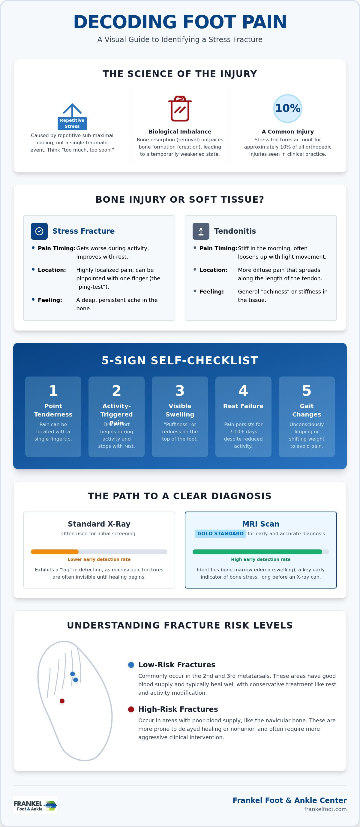
The Stress Fracture Checklist: 5 Signs You Should Not Ignore
Identifying a stress fracture in foot requires a shift in how you monitor physical discomfort. Because these injuries lack the dramatic onset of a "snap" or a sudden fall, they often masquerade as simple muscle soreness or fatigue. To help you determine if your pain requires clinical intervention, we have compiled a checklist based on the most common clinical presentations for symptoms and diagnosis of stress fractures. If you recognize these patterns, your body is likely signaling a structural issue that rest alone may not resolve.
- Point Tenderness: This is a major red flag. If you can pinpoint the exact source of your pain with a single finger, rather than feeling a general area of ache, bone involvement is highly probable.
- Activity-Triggered Pain: Pay attention to the timing. Does the discomfort start shortly after beginning a walk or run, but disappear almost immediately upon sitting down? This specific cycle is a hallmark of early bone stress.
- Visible Swelling: Examine the top of your foot for "puffiness" or redness over the metatarsal area. If you lose the visible definition of the tendons on one foot compared to the other, inflammatory fluid is present.
- Rest Failure: Most soft tissue strains improve within a few days. If the pain persists for more than 7 to 10 days despite significantly reducing your activity levels, professional screening is necessary.
- Gait Changes: Are you unconsciously shifting your weight to the outside of your foot or limping to avoid pressure on the sore area? Altering your stride can lead to secondary injuries in the ankle or knee.
When to Call a Specialist in Middletown, Katonah, or Milford
While some minor injuries respond to home care, certain high-risk areas demand urgent attention. The base of the fifth metatarsal and the navicular bone have poor blood supply, making them prone to non-union or complete breaks if left unsupported. If you cannot walk a full block without significant pain, it's vital to schedule a consultation at Frankel Foot & Ankle Center for a definitive diagnosis. Early intervention at our clinics in Middletown, Katonah, or Milford is the most effective way to ensure you don't face a prolonged recovery or permanent mobility issues.
Advanced Diagnostics and The Path to Pain-Free Mobility
One of the most frustrating aspects of a stress fracture in foot is the "X-ray lag." Patients often visit a clinic early in their pain journey only to be told their imaging appears normal. This occurs because standard X-rays frequently fail to show microscopic bone changes for the first 14 to 28 days. It is only after the body begins to form a "callus," or new bone growth to bridge the crack, that the injury becomes visible on traditional films. Relying solely on an early negative X-ray can lead to continued activity that worsens the injury.
Advanced diagnostics and treatment now rely on MRI as the clinical gold standard for early detection. An MRI is significantly more sensitive, allowing specialists to identify bone marrow edema, which is swelling inside the bone, before a physical crack even forms. This allows for earlier intervention and a more predictable recovery timeline. Once diagnosed, the primary goal shifts to offloading the affected area. This typically involves using stiff-soled shoes or specialized orthopedic boots to redirect weight away from the vulnerable bone, ensuring the environment is right for comprehensive foot pain relief and structural healing.
Modern Recovery Protocols: Beyond Just Rest
Recovery doesn't have to mean total inactivity. We utilize "active rest" protocols to keep you moving while your bone heals. This involves transitioning to low-impact activities such as swimming or stationary cycling, which maintain your cardiovascular fitness without placing repetitive loading forces on the stress fracture in foot. Additionally, we focus on nutritional optimization. Ensuring adequate levels of Calcium and Vitamin D is essential, as these nutrients provide the necessary biological building blocks for the bone remodeling process.
Preventing Future Foot Injuries
Preventing a recurrence is just as important as the initial healing phase. We recommend a structured "Return to Play" framework that follows the 10% rule: never increase your exercise duration, intensity, or mileage by more than 10% per week. To address the root cause of the stress, a professional gait analysis can identify biomechanical imbalances, such as overpronation or high arches, that may be overloading specific bones. For more information on maintaining lower extremity health, you can explore our Ankle Pain Relief pillar, which details how ankle stability impacts foot health.
Take the First Step Toward Lasting Mobility
Navigating the uncertainty of a stress fracture in foot doesn't have to be a solitary journey. By identifying the signs of point tenderness early and understanding why advanced imaging is essential for an accurate diagnosis, you've already taken the most important step toward protecting your long-term health. Proper recovery involves more than just resting; it requires a structured approach that balances active movement with clinical precision to prevent recurrent injuries and ensure your bones heal with the necessary structural integrity.
At Frankel Foot & Ankle Center, we specialize in providing sophisticated, minimally invasive podiatric solutions designed to restore your quality of life. Whether you're visiting our offices in Middletown, Katonah, or Milford, you'll receive expert care and a thorough diagnostic evaluation led by Dr. Marc Frankel. Don't let a "silent" injury dictate your future activity levels or lead to chronic complications. We're here to provide the definitive answers and personalized recovery protocols you need to move forward without pain.
Book an Appointment at Frankel Foot & Ankle Center today to secure your path to a safe and efficient recovery. You have the strength to heal, and we have the expertise to guide you every step of the way.
Frequently Asked Questions
Can I walk on a stress fracture in my foot?
Walking on a suspected stress fracture is generally discouraged until you have received a professional evaluation and a protective device. Continued weight-bearing can cause a microscopic crack to expand into a complete break, significantly complicating your recovery. While you may be able to tolerate the pain, doing so risks permanent structural damage. A specialist will typically recommend a stiff-soled shoe or walking boot to offload pressure during the initial healing phase.
How long does a foot stress fracture take to heal?
The typical recovery period for this injury is six to eight weeks of restricted activity. This timeline allows the bone remodeling process to successfully bridge the microscopic fissures with new, healthy bone tissue. Your specific duration may vary depending on the bone involved and your adherence to the treatment plan. High-risk locations, such as the fifth metatarsal, often require a more cautious approach to ensure the bone heals without the risk of non-union.
Will a stress fracture show up on an X-ray immediately?
No, a stress fracture in foot rarely appears on a standard X-ray during the first two to four weeks after the onset of pain. These injuries are often invisible until the body begins to form a "callus," or new bone growth, around the site of the injury. For this reason, we rely on advanced diagnostics like MRI to detect bone marrow edema and internal inflammation long before the fracture becomes visible on traditional imaging.
What happens if I ignore a stress fracture and keep running?
Ignoring the pain and continuing to run can transform a manageable injury into a chronic condition that requires surgical intervention. The repetitive stress prevents the bone from healing, which can lead to a complete fracture or a permanent deformity in the foot's structure. Furthermore, a neglected stress fracture in foot often causes you to alter your gait, leading to secondary injuries in your ankles, knees, and hips due to improper weight distribution.


